CONCISE ANATOMY
Where Anatomy
Meets Clarity
We make anatomy simple, visual, and unforgettable — empowering learners and educators worldwide.
A Comprehensive, Free Learning Resource
Our platform offers 780+ peer-reviewed articles covering the breadth of human anatomy, all available for free to support your learning journey.
Regional & Systemic Anatomy
Master the body's structure through both regional and systemic approaches.
Clinical Oriented Anatomy
Bridge the gap between anatomical knowledge and clinical practice.
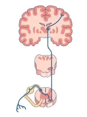
Neuroanatomy
Unravel the complexities of the nervous system's structure and function.
Embryology
Trace the developmental journey from a single cell.
CONCISE ANATOMY PREMIUM
An Exclusive Premium Experience in Our App
Unlock a complete learning ecosystem on your mobile device. All premium features are available exclusively in the Concise Anatomy App.
Image Atlas
Thousands of high-definition, labeled images and illustrations.
3D Models
Hundreds of interactive 3D models for a deeper understanding.
Flashcards
Test your knowledge with our extensive library of flashcard decks.
Videos
Engaging video lectures and 3D animations to simplify complex topics.
Podcasts
Learn on the go with our expert-led anatomy podcasts.
Question Bank
A personalized quiz platform with thousands of questions.
Download the App
Explore all premium features to study anatomy without limits.
Experience Anatomy Without Limits
Go beyond the basics with premium features — from precision visuals and realistic 3D models to smart quizzes and audio-guided lectures — designed to make anatomy clearer and more memorable than ever.
High-Definition Image Atlas
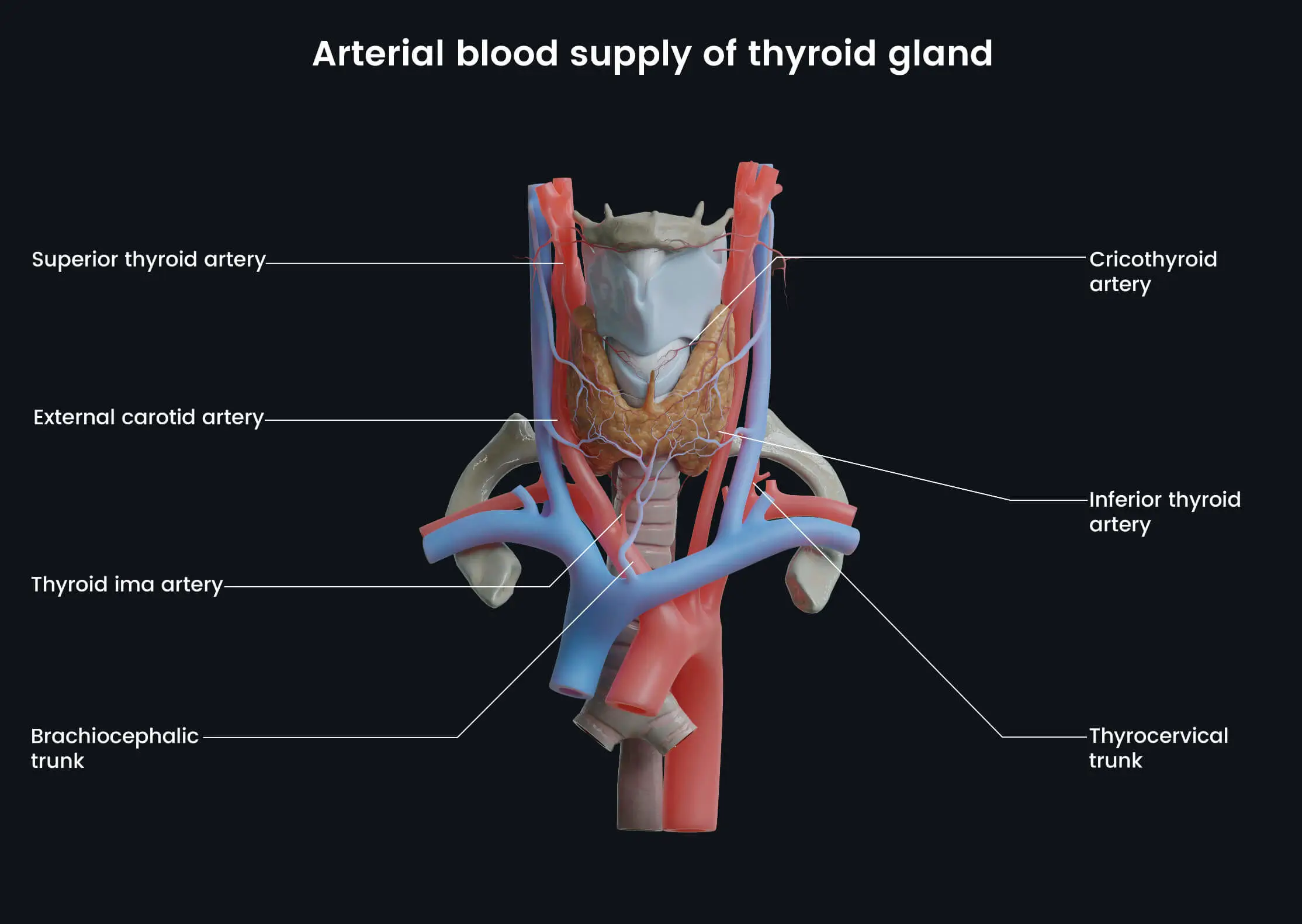
Thyroid Gland and Blood Supply
A highly vascular endocrine gland located in the anterior neck. This illustration highlights its rich arterial supply from the superior and inferior thyroid arteries and venous drainage via the superior, middle, and inferior thyroid veins. Note its close relationship with the trachea and larynx.
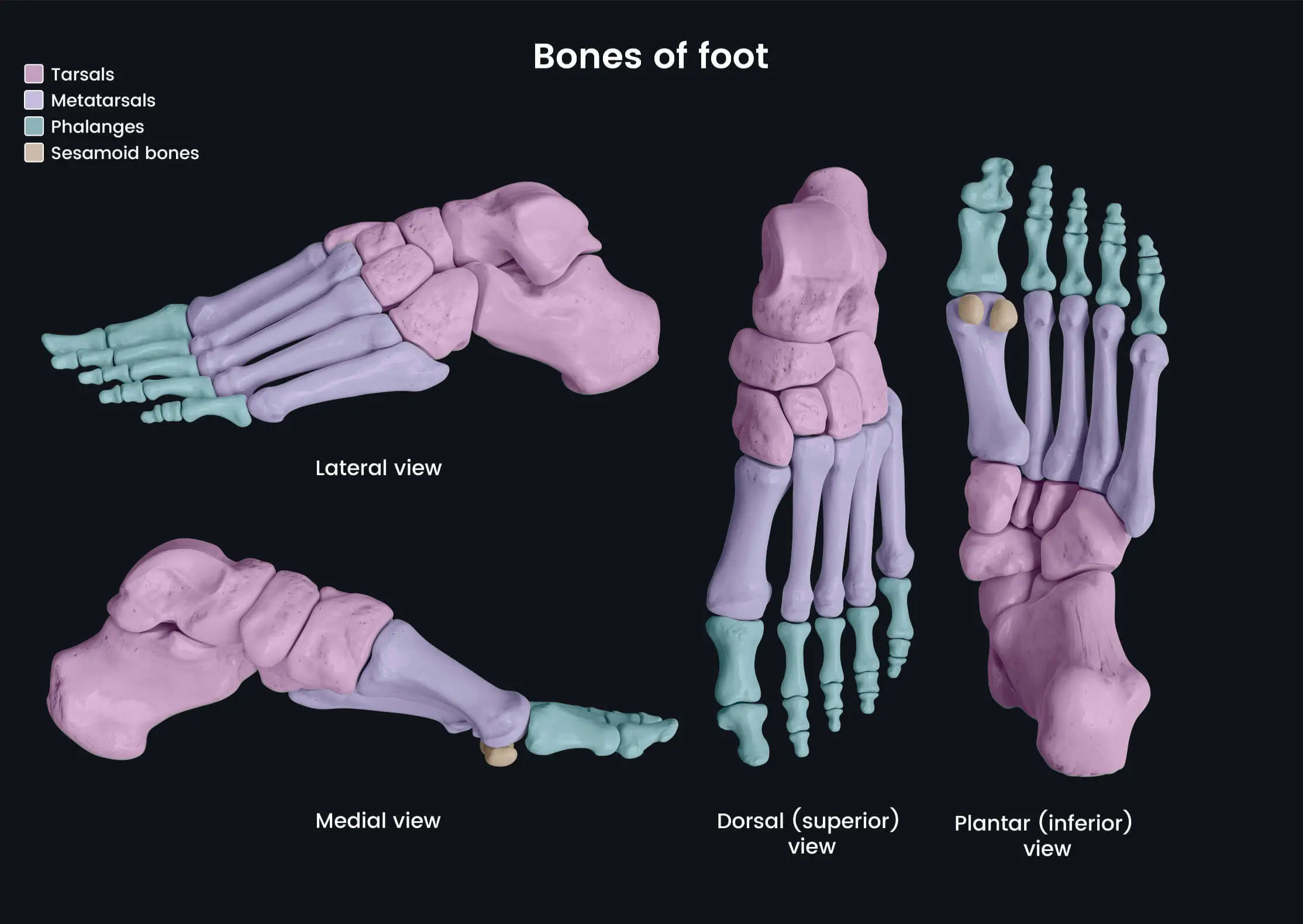
Bones of the Foot
The intricate structure of the foot, composed of 7 tarsals, 5 metatarsals, and 14 phalanges. They form the medial and lateral longitudinal and transverse arches, crucial for weight bearing and propulsion during gait.
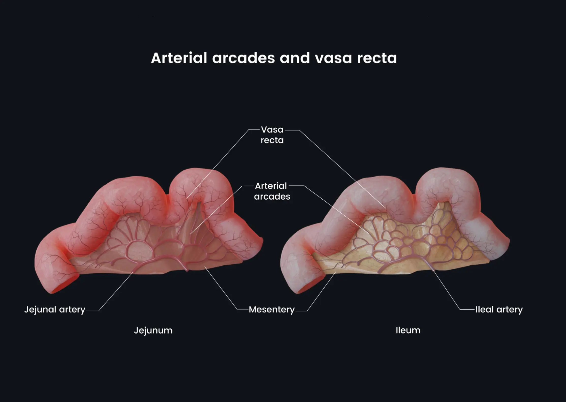
Arterial Arcades and Vasa Recta
Demonstrates the difference in blood supply between the jejunum and ileum. The jejunum has simple, large arterial arcades and long vasa recta, while the ileum has complex, shorter arcades and vasa recta, reflecting their differing absorptive functions.
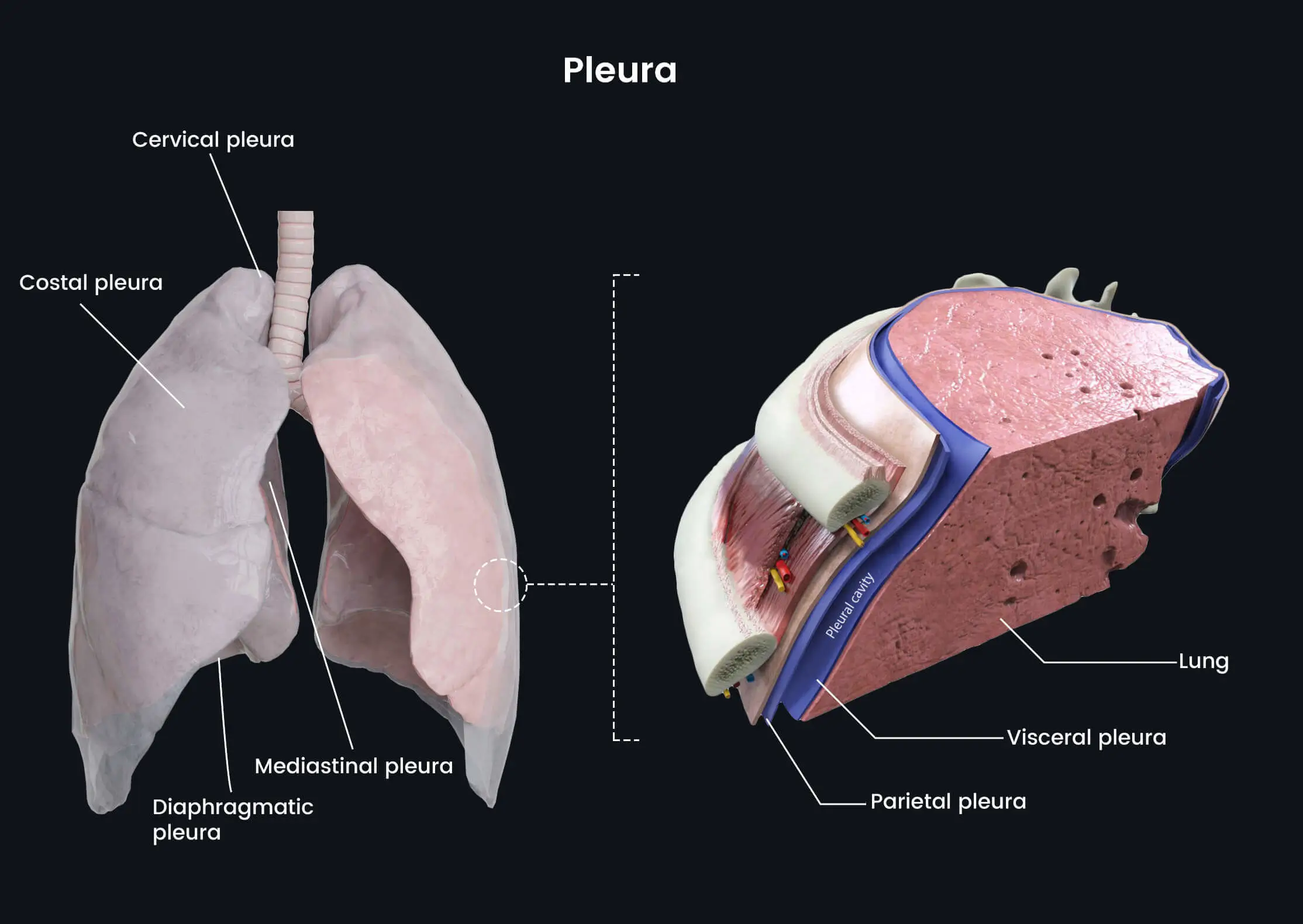
Lung, Pleura & Intercostal Neurovascular Bundle
A cross-sectional view showing the relationship between the lung, pleura, and thoracic wall. The intercostal vein, artery, and nerve (VAN) are shown protected in the costal groove between the internal and innermost intercostal muscles.
Explore all Images
Pre-Register for App
Immersive 3D Models
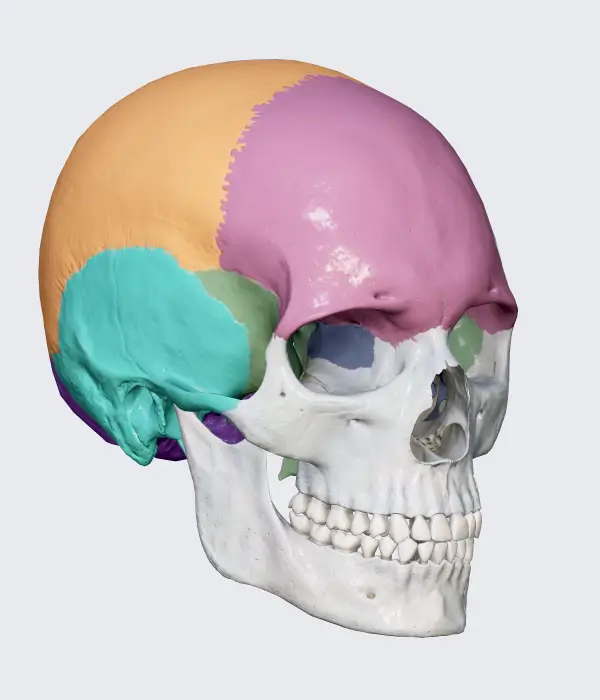
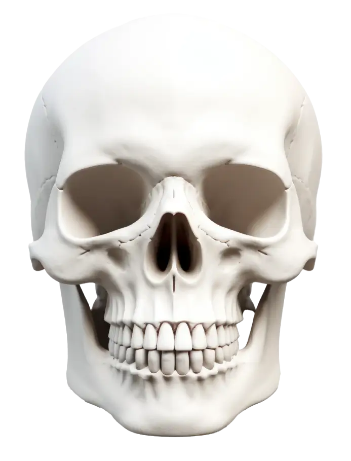
The Skull
Explore a richly detailed 3D representation of the human skull, allowing you to isolate individual bones and study their contours, articulations, and structural relationships. Examine the major cranial regions, view internal and external surfaces, and observe how neurovascular structures pass through key foramina such as the optic canal, jugular foramen, and foramen magnum. This expanded model also highlights sutures, sinus cavities, and clinically significant landmarks, giving you a deeper understanding of cranial organization, trauma patterns, surgical approaches, and anatomical variability.
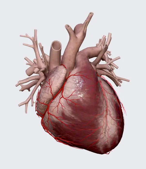
The Heart
Delve into a comprehensive 3D model capturing both the structural and functional aspects of the human heart. Navigate through chambers, valves, muscular walls, conduction pathways, and surrounding pericardial layers with smooth, precise visualization. Explore coronary arteries and veins in high detail, observe blood-flow routes, and understand the mechanical coordination of valve movement. This model also presents realistic relationships with great vessels, septal structures, and myocardial regions, making it ideal for mastering cardiac anatomy, imaging interpretation, physiology concepts, and clinical conditions such as ischemia or valve disorders.
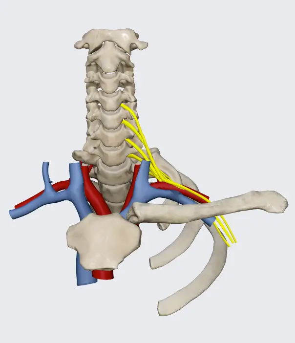
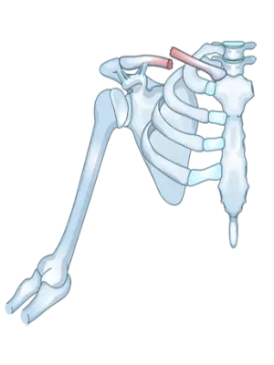
The Brachial Plexus
Visualize the entire brachial plexus in 3D, positioned accurately alongside key musculoskeletal and vascular structures of the neck, axilla, and upper limb. Trace each component—from roots to terminal branches—as they course around the scalene muscles, clavicle, first rib, and axillary artery. The model provides a clear, spatial understanding of nerve distribution, muscular innervation, and sensory territories. It also clarifies common injury mechanisms, anatomical variations, and clinically significant compression points, making an otherwise complex region intuitive and clinically meaningful.
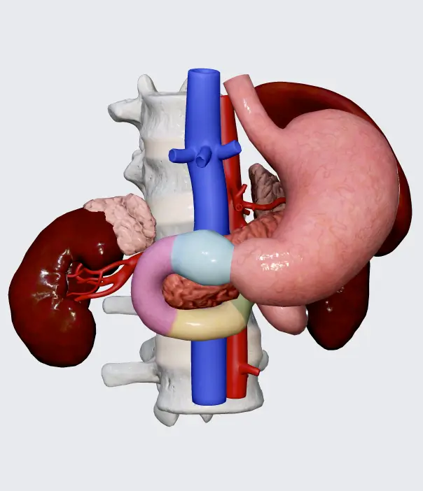
The Duodenum & Pancreas
Examine a highly realistic 3D reconstruction of the duodenum and pancreas, showcasing the full C-shaped duodenal curvature wrapped around the pancreatic head. Observe the spatial relationships between ducts, vessels, and surrounding retroperitoneal structures, including the bile duct, pancreatic duct, hepatic vessels, and major arterial arcades. This model highlights functional intersections such as the hepatopancreatic ampulla and provides a detailed appreciation of how disease processes—like ulcers, pancreatitis, and obstructive lesions—affect this densely packed anatomical region. Designed for comprehensive orientation, clinical insight, and advanced anatomical study.
Explore all 3D Models
Pre-Register for App
Learn with Videos & Animations
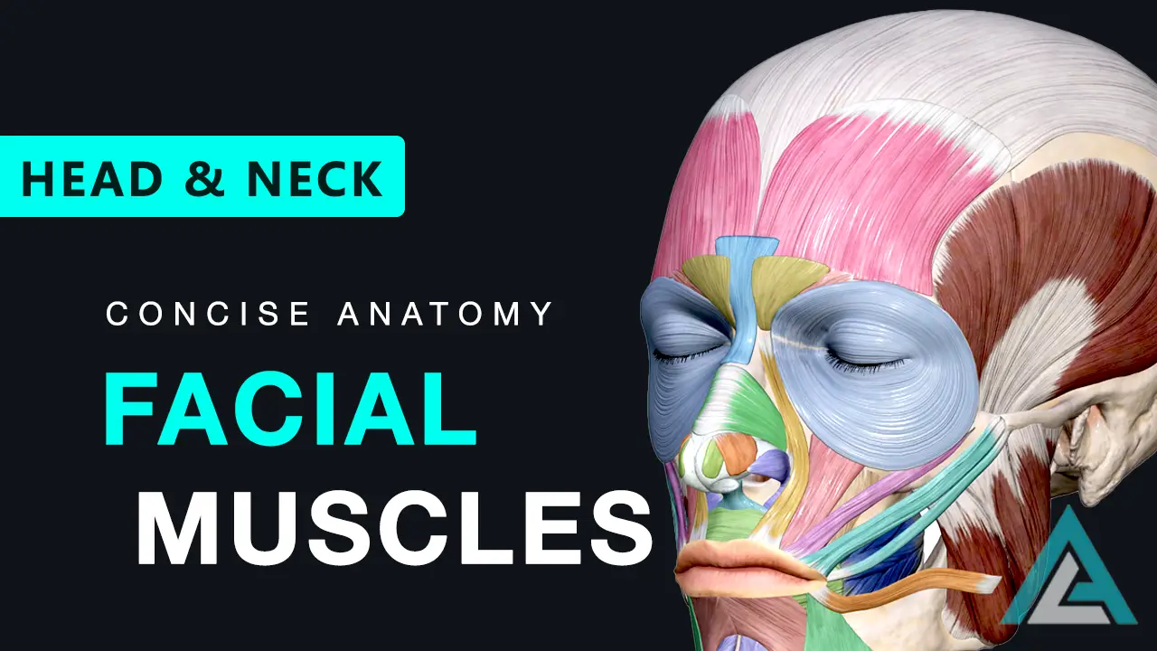
Muscles of Facial Expression
A comprehensive lecture on the structure and function of the facial muscles, crucial for expressions and emotions.
Humerus Bone Anatomy
A detailed guide covering the key features, muscle attachments, and clinical importance of the humerus.

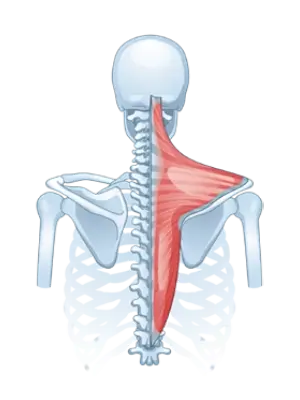
Muscles of the Back
A step-by-step exploration of the complex layers of the back, from superficial to deep intrinsic muscles.
Extraocular Muscles
Learn the anatomy and functions of the six muscles that control precise eye movements.
Listen to Clinical Anatomy Podcasts
Inguinal Canal Anatomy & Clinical Correlation
A breakdown of the walls, rings, and contents of the inguinal canal and its clinical link to hernias.
Thyroid Gland Anatomy & Clinical Correlation
An anatomical overview of the thyroid gland, covering its structure, relations, blood supply, development, and surgical considerations.
Upper Limb Deformities: Nerve Injuries
A clinical guide to upper limb nerve injuries, from brachial plexus lesions to Volkmann's and Dupuytren's contractures.
Explore all Podcasts
Pre-Register for App

Master Anatomy with Our Question Bank
Prepare smarter with our app-exclusive question bank — designed to strengthen your understanding and boost exam confidence.
- 2,800+ board-style questions with detailed explanations.
- Customizable quizzes by topic, region, or system.
- Smart performance tracking to identify your strengths and weaknesses.
Get full access in our Premium App, launching Early 2026. Try the web demo now!
Ready to Master Anatomy?
Join thousands of learners who trust Concise Anatomy for clear, visual, and unforgettable anatomy learning.