Overview
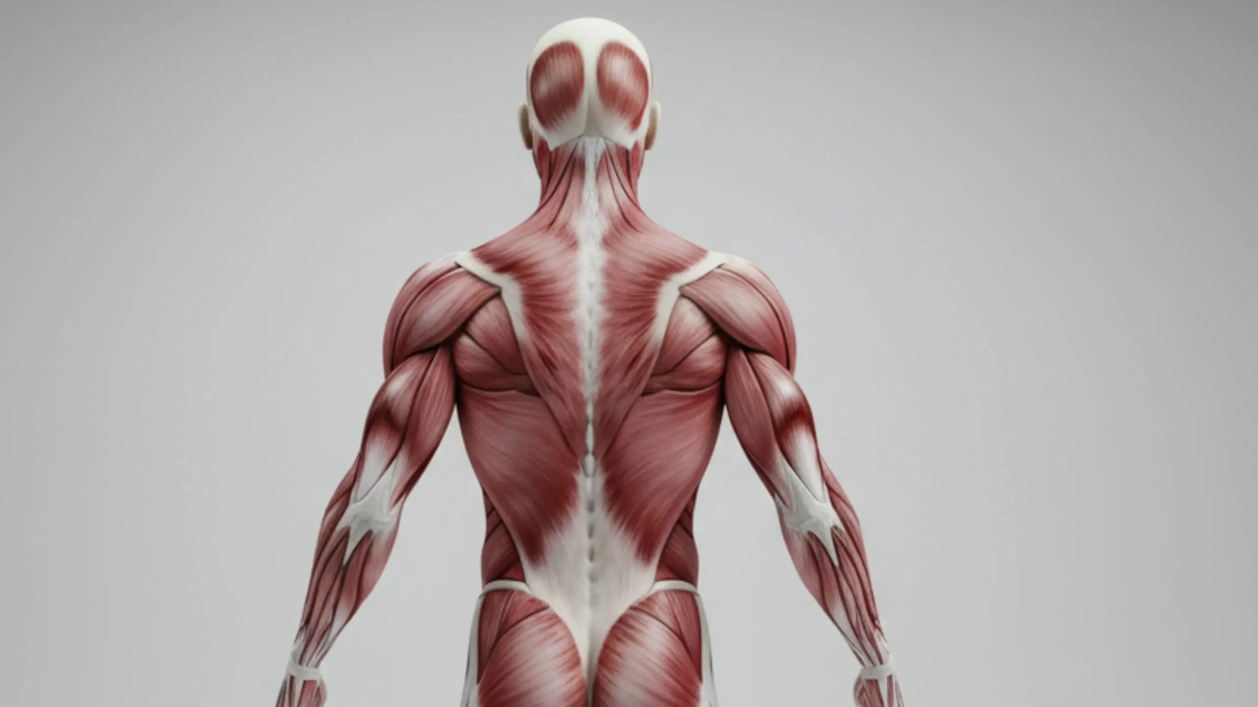
The muscles of the back form layered functional groups that span from the skin down to the vertebral column. Understanding these layers is crucial for interpreting posture, back pain, and movements of the upper limb. The superficial and intermediate groups are classically called extrinsic back muscles because they primarily move the limb or assist respiration, whereas the deep group consists of intrinsic back muscles that act directly on the vertebral column.
Before memorising individual muscles, it is useful to revise the bony framework of the vertebral column and general principles of skeletal muscle anatomy, then come back to this high-yield summary table.
For a quick visual reinforcement of the layered back anatomy, here’s a concise video walkthrough that pairs perfectly with the summary tables below.
Superficial (Extrinsic) Back Muscles
Superficial back muscles connect the axial skeleton to the pectoral girdle and humerus. They are supplied mainly by anterior rami via peripheral nerves, with the notable exception of trapezius, which is innervated by the spinal accessory nerve (CN XI). Functionally, they are more related to the shoulder region than to intrinsic back support.
Superficial Layer
| Muscle | Origin | Insertion | Nerve Supply | Main Action |
|---|---|---|---|---|
| Trapezius | External occipital protuberance; nuchal ligament; C7–T12 spinous processes | Lateral clavicle; acromion; spine of scapula | Spinal accessory nerve (CN XI); C3–C4 sensory | Upper fibres elevate scapula; middle fibres retract; lower fibres depress; upper + lower fibres rotate glenoid cavity upward |
| Latissimus Dorsi | T7–T12 spines; thoracolumbar fascia; iliac crest; inferior ribs | Floor of intertubercular sulcus of humerus | Thoracodorsal nerve (C6–C8) | Extends, adducts, and medially rotates humerus |
| Levator Scapulae | Transverse processes of C1–C4 | Superior angle and medial border of scapula | Dorsal scapular nerve (C5); cervical nerves (C3, C4) | Elevates scapula; rotates glenoid cavity downward |
| Rhomboid Major | T2–T5 spinous processes | Medial border of scapula (spine to inferior angle) | Dorsal scapular nerve (C4, C5) | Retracts and elevates scapula; assists downward rotation |
| Rhomboid Minor | Nuchal ligament; C7–T1 spinous processes | Medial border of scapula at spine level | Dorsal scapular nerve (C4, C5) | Retracts and stabilises scapula; marks surface landmark for spine of scapula |
Intermediate (Extrinsic) Back Muscles
The intermediate back muscles form a thin muscular layer overlying the intrinsic back muscles. They attach to the ribs and are thought to assist respiratory movements rather than major spinal actions. To understand their relationship to the ribs and intercostal spaces, revise the respiration mechanism and intrinsic muscles of the thoracic wall.
Serratus Posterior Group
| Muscle | Origin | Insertion | Nerve Supply | Main Action |
|---|---|---|---|---|
| Serratus Posterior Superior | Nuchal ligament; C7–T3 spinous processes | Ribs 2–5 (superior borders) | Intercostal nerves (T2–T5) | Elevates ribs; assists inspiration |
| Serratus Posterior Inferior | T11–L2 spinous processes | Ribs 9–12 (inferior borders) | Intercostal nerves (T9–T12) | Depresses or stabilises lower ribs |
Deep (Intrinsic) Back Muscles
Intrinsic back muscles span from the sacrum and pelvis to the skull and are enclosed by the deep fascia of the back, including the thoracolumbar fascia. They are supplied segmentally by the posterior rami of spinal nerves and act directly on the vertebral column to maintain posture and control finely graded movements.
Sacrospinalis (Erector Spinae) Muscles
The erector spinae form the principal longitudinal muscle mass of the back. From lateral to medial, they are arranged as iliocostalis, longissimus, and spinalis. Together they are the main extensors of the vertebral column and key postural muscles.
| Muscle | Origin | Insertion | Nerve Supply | Main Action |
|---|---|---|---|---|
| Iliocostalis (Lumborum, Thoracis, Cervicis) | Iliac crest; sacrum; lumbar fascia; spinous processes of lower lumbar vertebrae | Angles of ribs; cervical transverse processes | Posterior rami of spinal nerves | Extends and laterally flexes vertebral column; most powerful in lumbar and thoracic regions |
| Longissimus (Thoracis, Cervicis, Capitis) | Sacrum; lumbar vertebrae; thoracic transverse processes | Transverse processes; ribs; mastoid process of temporal bone | Posterior rami of spinal nerves | Extends vertebral column and head; lateral flexion; rotates head to the same side |
| Spinalis (Thoracis, Cervicis) | Spinous processes of upper lumbar and lower thoracic vertebrae | Spinous processes of upper thoracic and cervical vertebrae | Posterior rami of spinal nerves | Weak extensor of thoracic and cervical spine; often blends with semispinalis |
Transversospinal Muscles
Transversospinal muscles lie deep to the erector spinae, running obliquely from transverse to spinous processes. They provide fine control, stabilisation, and rotation of the vertebrae and are especially important in segmental spinal mechanics.
| Muscle | Origin | Insertion | Nerve Supply | Main Action |
|---|---|---|---|---|
| Semispinalis (Thoracis, Cervicis, Capitis) | Transverse processes of thoracic and lower cervical vertebrae | Spinous processes of cervical and thoracic vertebrae; occipital bone | Posterior rami of spinal nerves | Powerful extensor of head, neck, and thorax; causes contralateral rotation of vertebral column |
| Multifidus | Sacrum; ilium; mammillary and transverse processes | Spinous processes 2–4 segments above origin | Posterior rami of spinal nerves | Segmental stabiliser; important for lumbar spine stability |
| Rotatores (Brevis & Longus) | Transverse processes of vertebrae (most developed in thoracic region) | Lamina or spinous process 1–2 segments above origin | Posterior rami of spinal nerves | Fine-tunes rotation and stabilisation of adjacent vertebrae |
Minor Deep Layer
The minor deep muscles are short segmental muscles that bridge adjacent vertebrae or vertebrae and ribs. They assist the larger intrinsic back muscles in extension, lateral flexion, and subtle segmental control.
| Muscle | Origin | Insertion | Nerve Supply | Main Action |
|---|---|---|---|---|
| Interspinales | Spinous processes of cervical and lumbar vertebrae | Spinous process immediately above | Posterior rami of spinal nerves | Assist extension of vertebral column |
| Intertransversarii | Transverse processes of cervical and lumbar vertebrae | Transverse process immediately above | Anterior and posterior rami of spinal nerves | Lateral flexion of vertebral column |
| Levatores Costarum | Transverse processes of C7–T11 | Ribs below (between tubercle and angle) | Posterior rami of C8–T11 spinal nerves | Elevate ribs; assist respiration and lateral flexion of thoracic spine |
Suboccipital Region
At the craniovertebral junction, small intrinsic muscles form the suboccipital group. They bridge the atlas, axis, and occipital bone. For detailed regional relationships, see the suboccipital region and the broader muscles of the back of the neck pages.
Suboccipital Muscles
| Muscle | Origin | Insertion | Nerve Supply | Main Action |
|---|---|---|---|---|
| Rectus Capitis Posterior Major | Spinous process of C2 | Inferior nuchal line of occipital bone | Suboccipital nerve (posterior ramus of C1) | Extension and ipsilateral rotation of head at atlanto-occipital and atlanto-axial joints |
| Rectus Capitis Posterior Minor | Posterior tubercle of C1 | Medial part of inferior nuchal line | Suboccipital nerve | Assists extension of head; fine positional control |
| Obliquus Capitis Superior | Transverse process of C1 | Occipital bone between superior and inferior nuchal lines | Suboccipital nerve | Extension and lateral flexion of head |
| Obliquus Capitis Inferior | Spinous process of C2 | Transverse process of C1 | Suboccipital nerve | Rotates atlas on axis, turning face to same side |
Exam Strategy
For anatomy and clinical exams, first classify the back muscles by layer (superficial, intermediate, deep) and by function (upper limb movement, respiration, intrinsic spinal control). Then focus on patterns: intrinsic back muscles are supplied by posterior rami, extrinsic muscles by anterior rami, and suboccipital muscles by the C1 posterior ramus.
Use this summary alongside regional pages on the posterior abdominal wall muscles, vertebral canal, and spinal cord and tracts to integrate muscular anatomy with vertebral, neural, and fascial anatomy.